
When the Human Genome Project1 was launched in autumn 1990 with the aim of identifying and mapping all of the genes of the human genome, no-one would have thought that we would discover a new microcosmos revolving around and mingling with our human cells.
Of course, already long before this project, it was well-known that our body is not sterile and there are many bacteria living within and on it. These bacteria were however mainly classified as being malicious, threatening our health and causing problems.2 Until the 70s of the last century, a germ-free personal environment was considered as most desirable, and strong cleaning products became quite popular. Only starting in the early 1980s, these ideas and information were carefully reevaluated.
When the first results of the Human Microbiome Project3 that started in 2008 were published and we learnt that not only do we host an amazing amount of microbes in and on our body but that these tiny cohabitants at least equal or possibly even exceed the amount of our own human cells, intense research has been dedicated to understand their function for human health and disease.4
With the rapid development of novel metagenomic methods, microbiome analysis has become much easier, quicker, more comprehensive and reliable than with traditional techniques such as sampling and culturing that only allow the identification of certain isolated species that can be cultured.
Besides the composition of our gut microbiome, also the microbial colonization of our skin which serves as our outside border to the environment has become the subject of intense
research.5, 6
While research of microorganisms on the skin in the past was mainly serving the purpose of identifying pathogens that cause disease and studying the means to fight them, only recently, the beneficial aspects of our skin commensals were discovered and further studied.

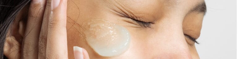
Figure 1: The skin is a natural habitat for a rich variety of microorganisms.
Many publications are describing their interaction to create a stable environment and protect their community and thereby also the skin from the invasion of foreign and potentially harmful microbes. Strong evidence of the interaction of the microbiome with the human immune system has been found.7 The microbiome seems to be involved even in protecting the body from UV radiation.8 On the other hand, many studies have proven that there is a close link between skin diseases such as acne9 and atopic dermatitis10 and the skin microbiome. Recent research indicates that “the microbiome is crucial for healthy skin, but so is healthy skin for the health of the microbiome” (Kristin Neumann11).
From these insights, we can conclude that by strengthening and balancing the microbial symbiosis, skin condition and health can be improved. Contrary, it seems clear that the use of antibiotics, strong disinfectants and cleaners may lead to a disruption of the microbial balance and impair skin health.
When the first evidence of the beneficial effects of our microbiome was found, it did not take long for more and more publications coming up about the possible improvement of the skin and its microbiome by pre-, pro- and postbiotic additions to cosmetic products.
Classic claims, such as the microbe-eliminating effects of cleansers, antibiotics and disinfectants can easily be proven, often by traditional methods focusing on individual species. Also, products that claim to leave the microbiome uninfluenced can be tested by metagenomic analysis.
Claims of balancing or improving the microbiome with dedicated products cannot be verified that easily. It seems that the marketers of such claims are far ahead of the scientists. Even though the most common bacteria on the skin have been identified, the microbial profiles of different individuals are significantly different.12 This is attributed to genetic and demographic properties, age, gender, ethnicity, skin type, lifestyle, hygiene, geographic differences, environmental stress by temperature, humidity, seasonal variation, radiation exposure and pollution, cohabitation with animals, profession and many more. Even applied products might cause shifts in their composition.
But not only between different people, even intra-individually, there is a huge variation of the skin conditions at different body sites.13 The face offers home to microbes that profit from or at least tolerate high skin surface moisture and increased sebum production. On the other hand, the extremities are rather dry and provide challenging conditions for most bacteria. The amount of microbes is much lower there than on the face, and a much higher variation between different persons has been found. Then, there are the arm pits and body folds with high moisture but moderate sebum content, again offering a completely different environment to microorganisms. And not only different moisture and sebum contents provide different microbial environments, there is also a huge impact of the skin pH. Bacteria regarded as beneficial usually prefer acidic conditions.
Because of all these influencing variables, until today there is no scientific consensus about the composition of the “ideal” microbiome. Microbiome analysis can show if the bacteria of probiotic products or the nutrition for special bacterial species in prebiotic formulations will lead to an undesired or a desired shift in the microbiome composition. Also, for some skin problems such as atopic dermatitis and acne, changes in the microbiome may be used to confirm the product efficacy. For cosmetic effects on healthy skin, however, it is almost impossible to interpret the result of such an examination.
If microbial analysis does not yield the desired information on the improvement of the skin condition, how else can we measure the efficacy of cosmetic claims for microbiome care products?
We have learnt that a healthy skin and a healthy microbiome are closely intertwined. Even though with our current knowledge, it is not possible to clearly quantify an improvement of the microbiome, skin health can be and has been measured for years. Different skin functions and conditions are ascribed to skin health.
Skin barrier assessment
The skin works as a barrier between our inner body and the environment. The strength of the skin barrier is closely linked to skin health: healthy skin will provide a strong barrier. Assessment of the barrier quality is usually performed by measuring the so-called TransEpidermal Water Loss (TEWL).14 This is the amount of water that evaporates through the skin into the environment. Healthy skin at most skin sites, measured at ideal ambient conditions of 20–22°C and 40–60% of relative humidity, will show a TEWL between 5–15 g/h/m2. If the barrier function is not optimal, a higher loss of water can be measured. Different probes for this application are available.

Figure 2: Tewameter®
measurement with open
measurement chamber does not influence the natural
evaporation from the skin.
Skin surface hydration measurement
The water holding capacity of the stratum corneum is depending on the presence of natural moisturizing factors and an intact skin barrier function. It is easily affected by many external influences such as dry air, absence of sebum, frequent washing etc. as this skin layer interacts strongly with the environment.
Dry skin will not only look scaly, but can also lead to small fissures in the skin surface where pathogens may settle. On the other hand, in a continually wet environment, the corneocytes that are forming the bricks in our skin wall will macerate which can also lead to a disruption of the skin integrity. The stratum corneum hydration therefore should be balanced, not too high but neither too low.
The water content will not only vary between persons with different skin types, but also between different skin sites on the single individual. At dryer skin sites like the arms less bacterial species can be found than in areas with higher moisture such as the face.
The Corneometer® is recognized throughout the industry as the gold standard to determine the stratum corneum hydration in the most important upper layers by capacitance. The measurement takes less than one second, thus not influencing the skin at all. Also, the L’Oréal Skinchip® or the MoistureMap® devices are based on capacitance measurement. They display the distribution of the water on the skin by capacitance imaging. Other equipment available on the market uses impedance or conductance methods.
Figure 3: The Corneometer® allows quick and easy hydration measurement at different skin sites.
Skin pH-value
Skin pH is essential for the well-being of specialized bacterial communities. Many bacteria associated with a healthy microbiome prefer acidic conditions and cooperate with the skin and amongst each other to maintain this microenvironment. Skin pH measurement can show if an applied product supports this aspect or if it leads to a shift of the pH value.
Figure 4: pH-changes
may influence microbial association. Their measurement can give valuable information on the efficacy of products.
From neutral pH values in the lower epidermis, there is a steep decrease of pH values towards the skin surface. Depending on the body site, age, gender and many other intrinsic and external factors, values at the skin surface mainly range between pH 4–6.15 Higher pH values at certain body sites like the axilla or intertriginous areas promote the colonization of certain odour-producing or harmful bacteria and fungi.16, 17
Cleaning the skin with soaps will lead to a short-term shift of the pH value. Even pure water with its neutral pH of 7.0 will influence the surface pH for a short period. Healthy skin will be able to balance this disruption within a certain time span. Frequent washing can however lead to cumulative effects and can seriously harm the skin barrier.18
Especially atopic dermatitis is often linked to increased pH-values and at the same time a different composition of the bacterial associations with an increased presence of staphylococcus aureus.19 Skin pH measurement in that case can show whether a shift of the pH value could be obtained by the product application.
Skin sebum
The Cambridge Dictionary describes the lipids excreted by the sebaceous glands as “an oil-like substance produced by the sebaceous glands in the skin that makes hair shiny and prevents skin from becoming dry”.20 The role of sebum in atopic dermatitis has been dismissed by different investigations in the past. More recent studies however, have shown that in diseased skin conditions such as atopic dermatitis, sebum levels are considerably lower than in healthy individuals.19 On the other hand, increased sebum production is correlated to the occurrence of acne.21

Figure 5: Sebum measurement with the Sebumeter® takes approximately
30 seconds.
Sebum is very different at different skin areas. In the face and on the scalp sebum levels are highest.22 Other skin sites show much lower values. The sebum content has a high impact on microbial life on the skin. Especially the facultative anaerobe cutibacterium acnes prefers sebum rich environments.
The Sebumeter® is the standard device to measure the sebum secretion on the skin surface. The special matte tape will become transparent by absorbing sebum present on the skin surface. The transparency will be measured – the higher, the higher the sebum level.
Other methods such as Sebufix® or Sebutape® use special microporous tapes that show the sebum pits on the skin surface as dark blotches of different sizes.
You can also study the presence of acne bacteria by using specialized camera equipment. The Cutibacterium acnes produces porphyrins that will show a fluorescence when exposed to UV light of special wavelengths.23 Even though these bacteria can be found on most people at oily skin sites, their activity poses a problem for acne-prone skin. Assessment of the porphyrin fluorescence is useful to test the efficacy of special anti-acne products. This can be done by using cameras providing the needed wavelengths. Special software will evaluate the number, area and intensity of fluorescence.

Figure 6: Strong fluorescence of the Cutibacterium acnes porphyrins under UV-illumination of the Visiopor® camera.
Figure 7: Homogeneous illumination and elimination of ambient light with the VisioFace® for standardized facial photography.
Evaluation of skin blemishes
Blemishes are usually an indication that the skin condition is not ideal. They especially occur in acne and are often correlated to an increased sebum production.
The occurrence of pimples and blackheads can be visually assessed and graded. This can be done based on direct inspection or for better comparison by the evaluation of high-resolution photographs.21 The comparability of photos is strongly influenced by ambient light not only in reference to the light colour and intensity but also to the angle of light. Additionally, the position of the face and the angle of photographing as well as the distance between camera and face will have a huge impact on the outcome.
Therefore, simple mobile phone cameras or even high-end consumer cameras are not the best-suited tools for taking such pictures.
There is dedicated equipment available on the market that provides a homogeneous illumination, sometimes even the use of different light sources and a fixed positioning of face and other body sites for taking standardized photos. With such equipment, very reproducible and comparable images can be taken that are evaluated either by subjective scoring or by automatic calculation of the size of the pores and the amount of blemishes.
Skin healing properties
Skin healing can be assessed by creating small wounds by punch biopsy, excision or damaging the skin in a different way. The extent of the wound and wound closure time can be assessed using visual methods.
An elegant solution not involving an open wound is the measurement of the TransEpidermal Water Loss (TEWL), already described above, after standardized repetitive skin stripping with special tapes such as Corneofix® or D-Squame®. The damage will show in an increased TEWL and can be evaluated together with the time the skin takes to recover to healthy TEWL values.
Desquamation
The skin continuously sheds dead skin cells from the outmost layer of the stratum corneum to make room for new cells growing from the base of the epidermis. This desquamation process is triggered by enzymatic activities that are induced in different skin layers, often depending on special pH values.25 Desquamation has been related to skin health and barrier function. In healthy skin, the shedding of corneocytes should be more of less imperceptible and very regular. When the desquamation process is disrupted, the shedding tends to become inhomogeneous, leaving islands of rather thick layers of dry corneocytes that can sometimes even be perceived by the eye as scaliness.
Desquamation is easily assessed by using the same type of tapes as for standardized tape stripping, e.g. the Corneofix® or the D-Squame®. These sticky tapes remove lose corneocytes. Combined with special cameras and evaluation software, you can quantify the degree of desquamation. In the resulting picture, accretions of corneocytes appear in whitish colour, whereas the regular desquamation will show in grey colours. Homogeneity or irregularities in desquamation can be easily displayed by using false colours.
Figure 8: Desquamation measurement with Corneofix® using Visioscan® camera and software – left side: irregular and strong desquamation, right side: fine and regular desquamation
Measurement of skin irritation
Skin irritation will show in increased microcirculation visible as an increased redness.24 Its intensity is often assessed by subjective scoring performed by experienced scientists. The human eye is, however, not able to evaluate very small differences and compare them to colours that have been previously seen at a different examination day. Photographs, taken at standardized conditions, will assist the researchers in the grading of redness.
Instead of subjective grading, image colour analysis can also be automatically performed by special software.
Besides comparing pictures, colour measurement to assess redness is often done by probes that are quicker and easier to use.
Also, other parameters such as skin surface topography or elasticity may offer information on skin health.
Conclusions
Research on the skin microbiome is an interesting and trendy topic that will probably evolve further. Despite the intense research of the last years, there is until now no scientific consensus on the perfect composition since the microbiome shows huge both inter- and intra-individual variation. Microbiotic analysis will therefore in most cases not yield satisfying prove of the efficacy of pre- and pro-biotic formulations for improved skin health.
You can easily prove skin health claims that are attributed to an improved microbiome by using traditional biophysical testing methods and imaging.
Some aspects that still need more research effort are the influence of cosmetic products on the resident microbiome. There are some concerns that microbiome influencing products may shift the scope of the microbiome by favouring only special microorganisms, thereby disturbing the balance of a perfectly healthy microbiome.
The role of environmental influences on the skin microbiome also offers a wide field of research.
These doubts need to be addressed in future tests and might even lead to the definition of new skin types, taking into account their main microorganisms.
References
- https://www.genome.gov/human-genome-project
- Howard I. Maibach und Gavin Hildick-Smith (Editors): Skin Bacteria and their Role in Infection, 1965, New York-Sydney-Toronto-London: McGraw-Hill Book Comp.
- NIH Human Microbiome Project, https://www.hmpdacc.org/overview/
- Baohong Wang, Mingfei Yao, Longxian Lv, Zongxin Ling, Lanjuan Li: The Human Microbiota in Health and Disease, Engineering, Volume 3, Issue 1, 2017, Pages 71-82, ISSN 2095-8099, https://doi.org/10.1016/J.ENG.2017.01.008. (https://www.sciencedirect.com/science/article/pii/S2095809917301492)
- Elizabeth A. Grice, Julie A. Segre: The skin microbiome, Nat. Rev. Microbio.l 9, 244–253 (2011). https://doi.org/10.1038/nrmicro2537
- Elizabeth A. Grice, Heidi H. Kong, Sean Conlan, Clayton B. Deming, Joie Davis, Alice C. Young, Gerard G. Bouffard, Robert W. Blakesley, Patrick R. Murray, Eric D. Green, Maria L. Turner, Julie A. Segre: Topographical and temporal diversity of the human skin microbiome, Science. 2009 May 29;324(5931):1190-2. doi: 10.1126/science.1171700. PMID: 19478181; PMCID: PMC2805064.
- James A. Sanford, Richard L. Gallo: Functions of the skin microbiota in health and disease. Semin. Immunol. 2013, Nov 30;25(5):370-7. doi: 10.1016/j.smim.2013.09.005. Epub 2013 Nov 20. PMID: 24268438; PMCID: PMC4219649.
- Vijay Kumar Patra, Karin Wagner,Velmurugesan Arulampalam, Peter Wolf: Skin Microbiome Modulates the Effect of Ultraviolet Radiation on Cellular Response and Immune Function, iScience 15, 211–222, May 31, 2019, https://doi.org/10.1016/ j.isci.2019.04.026
- Alan M. O’Neill, Richard L. Gallo: Host-microbiome interactions and recent progress into understanding the biology of acne vulgaris, Microbiome (2018) 6:177, https://doi.org/10.1186/s40168-018-0558-5
- Kazumasa Iwamoto, Masaya Moriwaki, Ryu Miyake, Michihiro Hide: Staphylococcus aureus in atopic dermatitis: Strain-specific cell wall proteins and skin immunity, Allergology International 68 (2019) 309-315.
- Panel Discussion: Panel II: Microbiome Mapping & Working with Skin Microflora, Moderator: Sarah Parsons (Editor) in Cosmetics Business, Dec. 2022, p. 24-25 and https://www.youtube.com/watch?v=jts0xPL7dcs
- E.K. Costello, C.L. Lauber, M. Hamady, N. Fierer, J.I. Gordon, R. Knight: Bacterial community variation in human body habitats across space and time, Science. 2009 Dec 18;326(5960):1694-7. doi: 10.1126/science.1177486. Epub 2009 Nov 5.
- B. Dreno, E. Araviiskaia, E. Berardesca, G. Gontijo, M. Sanchez Viera, L.F. Xiang, R. Martin,T. Bieber: Microbiome in healthy skin, update for dermatologists, JEADV, 2016, 30, 2038–2047, DOI: 10.1111/jdv.13965.
- J. Kottner: Transepidermal Water Loss in Young and Aged Healthy Humans: A Systematic Review and Meta-Analysis, Arch. Dermatol. Res., 23 January 2013, Volume 305: Pages 315-323, DOI: 10.1007/s00403-012-1313-6.
- Rippke F et al: The acidic milieu of the horny layer: New findings on the physiology and pathophysiology of skin pH. Am J Clin Dermatol 2002, Volume 3: Pages 261–272.
- Korting HC et al: Mikrobielle Flora und Geruch der menschlichen Haut. Hautarzt 1988; Volume 39: Pages 564–568.
- Stenzaly-Achtert S et al: Axillary pH and influence of deodorants. Skin Research & Technology, 2000, Volume 6: Pages 87–91.
- Strunk M et al: Regeneration des physiologischen Hautoberflächen-pH nach Anwendung von Hautreinigungsmitteln, Poster, 13. Dermatologisches Alpenseminar Grainau, 2018
- P.G. Sator et al: Comparison of Epidermal Hydration and Skin Surface Lipids in Healthy Individuals and in Patients with Atopic Dermatitis, J. Am. Acad. Dermatol., 2003, Volume 48: Pages 352-358; DOI: 10.1067/mjd.2003.105
- https://dictionary.cambridge.org/de/worterbuch/englisch/sebum
- G. Lanzendörfer, C. Uhl: How Effective is Your Anti-Acne Product, SPC, December 2018, Volume 91 (12): Pages 64-66
- J. Fluhr et al: Full Body-Skin Mapping for Six Biophysical Parameters: baseline Values at 16 Anatomical Sites in 125 Human Subjects, Skin Pharmacology & Physiology, 2012, Volume 25: Pages 25-33, DOI: 10.1159/000330721
- H. Dobrev: Fluorescence Diagnostic in Patients with Acne, Photodermatol. Photoimmunol. Photomed., December 2010, Volume 26(6): Pages 285-289. DOI: 10.1111/j.1600-0781.2010.00541.x.
- J.E. Wahlberg, M. Lindberg: Assessment of Skin Blood Flow – An Overview, in Bioengineering of the Skin: Cutaneous Blood Flow and Erythema (Berardesca E, Ed.), 1995, Pages 23-27
- Torbjörn Egelrud: Desquamation in the Stratum Corneum, Acta Derm. Venereol. 2000; Supp. 208: 44-45
Christiane Uhl
Christiane Uhl is Sales-, Project- and Training Manager of Courage + Khazaka electronic GmbH Cologne, Germany.
Fields of activity: Dermatological measurements, Scientific projects, Skin physiology, Educator skin testing